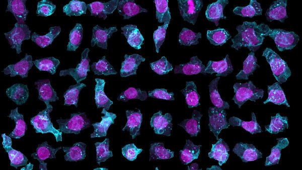
SEATTLE — For decades, peering inside a human cell has been a game of shadows, with microscopes and stains offering only fragmented glimpses that often damage the very structures they seek to reveal. Now, researchers at the Allen Institute have harnessed artificial intelligence to construct the first complete three-dimensional models of human cells, providing a detailed view of how cellular machinery rearranges itself in the face of disease.
The models, built from high-resolution scans of thousands of cells, depict a typical human induced pluripotent stem cell (hiPSC), chosen because the team was already cloning these cells to tag internal structures. The AI was trained on detailed images from 32,000 cells, and the resulting model can predict the location and arrangement of organelles—the specialized compartments within a cell—based on a single microscope image.
Beyond static visualization, the researchers also developed a probabilistic model that estimates organelle positions given a cell's size, shape, and nucleus location. This predictive capability could allow scientists to simulate how a cell's internal geography shifts under pathological conditions, such as cancer, without the need for invasive procedures.
Implications for Personalized Medicine
The practical payoff, according to the Allen Institute team, is a more nuanced understanding of how diseases alter cellular architecture. By feeding the AI images of cancerous cells, doctors could obtain a fuller picture of the disease's impact at the cellular level, potentially guiding treatment choices tailored to an individual patient's condition.
This approach addresses a long-standing limitation in medical imaging: traditional methods like stains and electron microscopy can alter or destroy the delicate structures they are meant to observe. The AI model offers a non-destructive alternative, generating a complete view from a single, minimally invasive image.
The team also hopes the tools will help democratize medical research, particularly in underserved regions where advanced imaging equipment may be scarce. By making the models freely available online, they aim to give researchers worldwide access to a level of cellular detail previously reserved for well-funded laboratories.
Looking ahead, the Allen Institute is working to expand its database, with plans to release models of additional cell types over the coming months. This ongoing effort promises to deepen our understanding of cellular behavior and open new avenues for disease diagnosis and treatment.



















Leave a Comment